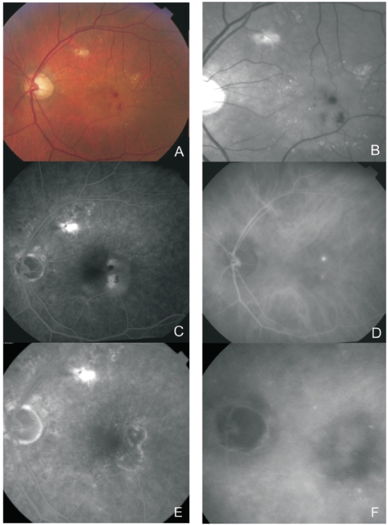
Figure 3 - RAP lesion.
Figure 3 - RAP lesion. Fundus colour photography (A) with intra-retinal haemorrhages, hard exudates and neurosensory detachment.
Red-free (B) with two juxtafoveal and extrafoveal small haemorrhages.
Fluorescein angiography (C) shows two juxtafoveal hyperfluorescent spots, neurosensory detachment and pigment epithelium detachment.
Late ICG reveals two juxtafoveal hyperfluorescent hot spots.
E and F: Fluorescein angiography and ICG after laser photocoagulation with resolution of exudation.