Figure 1 - RAP lesion
Figure 1 - RAP lesion
English
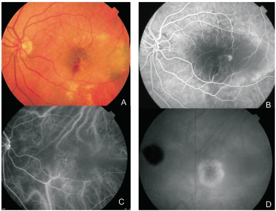
Figure 1 - RAP lesion. A: Fundus colour photography (A) with intra-retinal haemorrhages, hard exudates and neurosensory detachment.
Fluorescein angiography shows a retinal hiperfluorescent spot apparently at the end of one retinal vessel, an intra-retinal haemorrhage, neurosensory detachment and pigment epithelium detachment.
Early phase of ICG (C) with an intra-retinal hot spot (angiomatous proliferation) over diffuse choroidal hyperfluorescence.
Late phase ICG (D) reveals a subfoveal hiperfluorescent plaque (subretinal neovascularization).