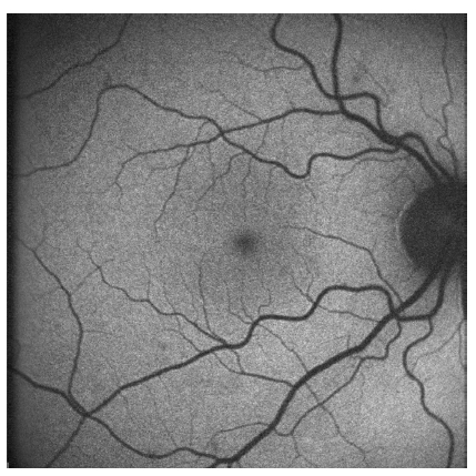
Figure 3: Normal fundus autofluorescence: uniform grayish signal in the fundus and a marked dark appearance in the optic nerve (absence of RPE) and retinal vessels (absorption of fluorescence by hemoglobin and other blood contents)
Figure 3: Normal fundus autofluorescence: uniform grayish signal in the fundus and a marked dark appearance in the optic nerve (absence of RPE) and retinal vessels (absorption of fluorescence by hemoglobin and other blood contents)
English
Figure 3: Normal fundus autofluorescence: uniform grayish signal in the fundus and a marked dark appearance in the optic nerve (absence of RPE) and retinal vessels (absorption of fluorescence by hemoglobin and other blood contents)